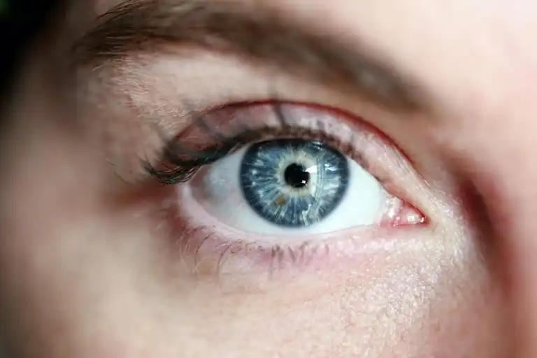
Imagine that your vision is the most advanced camera in the world. The retina is its film – an incredibly thin layer that captures light and transforms it into the images of your everyday life. Unfortunately, this precise structure can be unpredictable. Its damage is one of those moments in life when the fear of darkness becomes very real. At Swisslaser, however, we prove every day that modern medicine can work miracles, provided we act in time. Have you ever wondered whether retinal detachment is a death sentence? The answer is: absolutely not, but you need to know what to watch for.
What will you learn from the text below?
- What exactly is retinal detachment?It is a condition in which the retina loses contact with its base, cutting it off from its “fuel” – oxygen and nutrients.
- What symptoms should immediately concern you?If you see sudden flashes, floaters, or a dark “curtain” in front of your eye, do not delay for a minute.
- Who is at the greatest risk?Seniors, people with high myopia, and fans of contact sports should be particularly vigilant.
- Are there effective treatment methods for this condition?Yes, surgeons have vitrectomy and laser treatment at their disposal, which can effectively “reattach” the retina to its place.
- How can retinal detachment be prevented?Through regular examinations, we can identify weak points and secure them with laser before something serious happens.
Structure and Role of the Retina – Why Is It the Most Important Part of the Eye?
The retina is a true miracle of nature. Though thin as tissue paper, it consists of ten layers that work together with the precision of a Swiss watch.
The 10 Layers of the Retina: From Rods and Cones to the Optic Nerve
Inside the retina, millions of tiny workers – photoreceptors – are at work. You have rods that allow you to see in the dark, and cones that enable you to distinguish the blue of the sky from the green of grass. All of them collect signals and transmit them to the optic nerve, the great highway leading straight to your brain.
The Macula: The Center of Your Sharpest Vision
Did you know that reading fine print is the responsibility of just one small spot at the back of your eye? That is the macula. This is where the concentration of cones is greatest, allowing you to perceive the finest details on the face of a loved one.
How Does Light Turn into an Image? The Role of Visual Pigments
Pure chemistry takes place in your eye. Light strikes the visual pigments, causing them to break down, which in turn triggers an electrical impulse. It is remarkable that in a fraction of a second, your brain interprets these flashes as a complete image of the world.
Retinal Detachment – The Silent Threat to Your Vision
Unfortunately, retinal problems can strike unexpectedly, often without pain, making them extremely insidious.
What Is Rhegmatogenous Detachment and How to Recognize It?
Imagine that the vitreous body in your eye begins to shrink and pull on the retina. If it pulls too hard, a hole forms. Fluid then seeps underneath and begins to detach the retina from the wall of the eye – exactly like leaking water ruins wallpaper in a living room.
Alarming Symptoms: Flashes, Floaters, and a “Curtain” in Front of the Eye
This is the moment for a “red alert.” Do you see sudden flashes (photopsia), numerous new “cobwebs,” or a dark shadow approaching from the side? This is a signal to seek specialist help as quickly as possible. Time is your most valuable ally in this case.
Risk Factors: Age, Trauma, and Ophthalmic Surgery
Who should be most vigilant? Statistics point to men over sixty, but the risk also increases after cataract surgery or head trauma. If you are myopic, you should also have your fundus examined regularly!
Types of Detachment: Not Every Case Is the Same
Medicine also distinguishes other forms that are less commonly discussed but require completely different treatment.
Tractional Detachment in Diabetes and Retinopathy
Here, the culprits are scars and membranes that form, for example, in diabetes. They begin to “pull” on the retina, distorting the image. Straight lines suddenly become wavy – sounds alarming, doesn’t it?
Exudative Detachment: The Result of Inflammation and Coats’ Disease
In this version, the eye “sweats” from the inside. Fluid accumulates under the retina due to inflammation or tumors, pushing it out of its natural place. This is a rarer but no less serious situation.
Myopia and the Risk of Retinal Detachment
A long eyeball in a myopic person means that the retina is highly stretched. As a result, it is thinner and much more susceptible to damage during significant exertion. This is why examinations before planned childbirth are so important.
Modern Methods of Saving Vision
At Swisslaser, we fight for every eye, using technologies that until recently sounded like science fiction.
Vitrectomy and Tamponade: How Do Surgeons “Press” the Retina?
We remove the altered vitreous body and replace it with a special gas or oil. This “internal dressing” presses the retina against the choroid, giving it a chance to reattach.
Scleral Buckling: Stabilizing the Layers of the Eye from the Outside
This is a classic method in which we sew a tiny sponge onto the eye. Its task is to gently indent the wall of the eye inward so that it meets the detached retina. Effective and proven.
Laser Therapy and “Welding” the Retina to the Choroid
The laser is our most precise brush. We “weld” the edges of the tear with it, creating a permanent bond. This way, the fluid no longer has a path to further damage the structure of your eye.
Pregnancy and Retinal Condition: When Is a Cesarean Section Necessary?
Many expectant mothers worry about natural childbirth. The key to peace of mind is a visit to an ophthalmologist and a fundus examination.
Degenerative Changes and Natural Childbirth
If the retina has weak spots, the spike in pressure during pushing can be risky. The good news? Often, prophylactic laser treatment of weaker points is sufficient to allow safe natural childbirth.
Indications for Planned Cesarean Section: From Hemorrhages to Neovascularization
However, there are specific medical situations, such as active hemorrhages or detachment progressing during pregnancy. In such cases, your safety and that of your vision become a priority, prompting doctors to opt for a cesarean section.
Prevention and Protection: Laser Photocoagulation of the Retina
It is best to prevent a fire rather than extinguish it. Photocoagulation is our main preventive weapon.
What Does Laser Eye Protection Involve?
The procedure is quick, performed while sitting, and – most importantly for many patients – completely painless. We simply strengthen the retina in areas that appear suspicious.
Benefits of Photocoagulation: Reduction of Edema and Ischemia
With the laser, we seal leaking vessels and eliminate edema. This is the simplest way to avoid complicated surgeries in the future and enjoy peace of mind.
FAQ – Frequently asked questions
1. Does retinal detachment hurt?
That is the worst part – it usually does not hurt. If it did, patients would seek help sooner. The symptoms are purely visual.
2. Will I see the same as before after surgery?
It depends on how large an area detached and whether it involved the macula. A prompt procedure offers a tremendous chance of returning to full function.
3. Is laser treatment of the retina during pregnancy safe for the baby?
Absolutely. It is a local procedure that protects the mother’s vision and has no impact on the baby.
4. Do I have to lie in a specific position after vitrectomy?
Often, yes. If there is gas in the eye, we will ask you to keep your head in a specific position so that the gas bubble presses the retina where needed.
At Swisslaser in Warsaw and Krakow, we know how precious the gift of vision is. If you notice anything concerning – do not delay. Visit us and give yourself a chance for a clear tomorrow.
Do Not Delay Your Examination
Symptoms of retinal detachment require urgent diagnostics. We perform retinal imaging examinations.
- Fundus Examination — retinal assessment
- OCT Examination — imaging of retinal layers
- Contact — Krakow — tel. +48 12 383 33 60
- Contact — Warsaw — tel. +48 22 120 16 90
Sources
- Flaxel CJ, Adelman RA, Bailey ST et al. Posterior Vitreous Detachment, Retinal Breaks, and Lattice Degeneration Preferred Practice Pattern. Ophthalmology 2020;127(1):P146–P181. doi:10.1016/j.ophtha.2019.09.027
- Mitry D, Charteris DG, Fleck BW et al. The epidemiology of rhegmatogenous retinal detachment: geographical variation and clinical associations. Br J Ophthalmol 2010;94(6):678–684. doi:10.1136/bjo.2009.157727