Keratoconus at a glance
- Keratoconus is a progressive thinning and bulging of the cornea – it usually begins between the ages of 15 and 30 and is often mistaken for ordinary astigmatism at first.
- Warning signs: frequent changes in the cylinder of your glasses prescription, worsening vision at dusk, and distortions or ghost images despite a new correction.
- Modern treatment stops the disease: cross-linking (CXL) 3,600 zł per eye, CXL + laser corneal regularisation (the so-called Athens protocol) 8,700 zł, and CAIRS ring segments 16,000 zł per eye.
- The earlier keratoconus is detected, the simpler and less expensive the treatment – corneal tomography (PL) is the key.
- Full clinical guide: Keratoconus – symptoms, diagnosis and treatment.
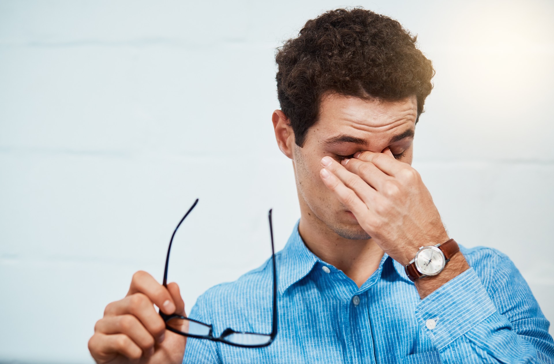
It’s not just people who work at a computer for long periods of time who often rub their eyes. If we were to pay attention to this trivial gesture, we would find that most of us have a habit of reaching into our eye and rubbing it whenever our eye starts burning and we feel sand under our eyelids.
This habit is detrimental to the eye. There’s a reason why we point out to children that they “shouldn’t push their paws into their eyes.” It is not only about the possibility of infecting the eye with pathogens and causing conjunctivitis, for example, but also about the increased risk of developing a corneal cone .
Corneal cone is a progressive eye disease that leads to thinning and distortion of the cornea, with subsequent deterioration of vision – myopia and astigmatism and often hypersensitivity to light.
Excessive habitual eyelid rubbing causes damage to corneal collagen. The middle layer of the cornea, the place where it is thinnest, bulges out under the pressure of the eye precisely into a cone shape.
Don’t push your paws into the eye!
Of course, not everyone who rubs their eyelids will develop corneal cone, although it is important to be aware that this is one of the causes of its formation. It is also worth remembering that corneal conditions are often accompanied by atopy or allergy, and then rubbing the eyelids is the answer to persistent itching.
Since uncontrollable rubbing of the eyelids always appears as one of the symptoms accompanying corneal cone – either as a cause or as an effect of already existing cone (yes, one can venture to say that the eye in cone is more itchy!), or the effect of accompanying allergic diseases – it is worth paying attention to other complaints besides itchy eyes, which can be treated as the first symptoms of corneal cone.
Failed again to select eyeglasses? Think about diagnostics for corneal cone!
Initially, the symptoms of corneal cone can be nonspecific – progressive deterioration of vision, blurring of vision, hypersensitivity to light, burning and redness of the eyes – so it is easy to underestimate the ongoing disease process.
Normally we go to the ophthalmologist, the latter selects glasses, but our eyesight still leaves much to be desired. So another visit, another pair of glasses and… nothing. We change ophthalmologist, blaming him for the failure of the selection of glasses and … still nothing.
We are beginning to get the impression that all around us are incompetent ophthalmologists who simply don’t know how to choose the right glasses for our vision defect. It happens that doctors order further tests and, paradoxically, thus prolong the correct diagnosis. Why? Because the very first wrong choice of glasses should raise suspicion that perhaps our problem is related to the fact that the deterioration of vision affects only one eye!
After all, we do most of our activities with both eyes during the day, and when vision deteriorates in one eye, we can often miss it. Only when the corneal cone also appears on the other eye do we fully realize that we see worse on both eyes.
As long as one eye sees badly, the selection of eyeglasses for the two eyes is therefore very difficult.
Corneal topography
To ascertain whether we are dealing with corneal cone one needs to perform corneal topography. Not every ophthalmologist’s or optometrist’s office has the right equipment, and in addition, the vision loss associated with high astigmatism during vision correction can behave very similarly to corneal cone and is sometimes mistaken for it.
What symptoms, besides itchy eyes, to watch out for?
We already know: itching and rubbing of the eyes, hypersensitivity to light, problems with the selection of eyeglasses – but what else should arouse our vigilance? Just mentioned symptoms associated with only one eye (monocular), such as:
- multiple vision – triple, quadruple vision
- distorted – and not necessarily only in one direction! – image
- halo, glare, starbust interference
- Afterimages, or so-called. ghost images.
Importantly, vision deteriorates to a similar degree to far and near distances, and the change in the quality of vision occurs rapidly over a very short period of time.
It also becomes troublesome to see after dark – so called. Twilight and night vision.
If you observe the symptoms listed above in yourself, or suspect that the trouble with your eyeglasses may be related to a developing corneal cone, do not delay in going to a good ophthalmologist who has the appropriate equipment.
Corneal topography is a non-invasive and painless examination. Depending on the changes that reveal in the study , measures are taken to:
- Zatrzymanie progresji – a w niektórych przypadkach cofnięcie – zmian chorobowych,
- The aim is for the patient to achieve the best possible visual acuity. If the cornea bulges only slightly, soft contact lenses are used; if the bulge is large, rigid lenses come into play.
Stopping the progression of corneal cone is paramount. Why? Because in the acute stages of the disease, the inner limiting membrane (Descemet’s) is torn, and the aqueous fluid from the anterior chamber of the eye enters the cornea itself.
We are then talking about the so-called. hydrops. The cornea then loses its translucency and a corneal transplant is usually required. Corneal cone can be intermediate or very advanced, reaching scores ranging from forme fruste keratoconus (preclinical stage) to grade IV cone.
At the SwissLaser Clinic, we perform CROSS-LINKING (named after the effect it produces) – also known as cross-linking of bonds, cross-collagenization of the cornea, or CXL – which causes the cornea to stiffen and increase its mechanical resistance. The stiffening of the cornea is accomplished by increasing the number of bonds between the collagen fibers that build it. It occurs under the influence of UVA irradiation of the cornea.
In order to increase the sensitivity of the superficial layers of the cornea to UVA radiation, it is soaked in a special photosensitizing substance, riboflavin (vitamin B2).
Clinical studies confirm that the action of riboflavin contributes to slowing or stopping the disease and, in some cases, its regression. Thus, the procedure makes it possible to bypass corneal transplantation.
I learned corneal surgery from Prof. Theo Seiler, co-inventor of the cross-linking method. The most important thing I can tell young patients with keratoconus is this: caught early, the disease remains a minor episode. Detected late, it can cost you your profession, your driving licence and your peace of mind. That is why every rapidly progressing change in cylinder deserves corneal tomography.
dr med. Victor Derhartunian, FEBO, FWCRS
Next step: book a qualification examination (400 zł, Warsaw or Kraków) – or first check the full keratoconus treatment price list.
Content medically reviewed by Dr. med. Victor Derhartunian, FEBO, FWCRS · Last updated: 17.08.2026