CATARACT TREATMENT IN CATOVICE

Choć najczęściej kojarzymy zaćmę (łac. cataracta) z podeszłym wiekiem pacjenta, może się ona pojawić także u dzieci. Congenital cataracts appear as early as birth, and can also develop in children during the first years of life. It arises as a result of metabolic (e.g. galactosemia) or chromosomal aberrations (e.g. Down syndrome), ocular malformations (lack of iris, microphthalmia, retinopathy of prematurity and others), hereditary conditions, intrauterine infections (e.g. when a pregnant woman becomes ill with rubella, hepatitis, toxoplasmosis, herpes and even influenza). Congenital cataracts can also be caused by fetal exposure to certain drugs in the womb (such as corticosteroids or sulfonamides). It may not give any spectacular symptoms, but sometimes a white pupil (Latin: leucocoria) can be noticed in a child. Leukocoria, also known as cat’s eye or white pupillary reflex, can appear not only in the course of congenital total cataracts, but also in the course of other eye diseases, such as a cancer called retinoblastoma, so noticing it absolutely requires an ophthalmologist’s consultation. Other symptoms suggestive of congenital cataracts include Franceschetti’s finger-eye reflex, which is the tendency to press the eye frequently with a fist or fingers and to put objects against the eye – because of the trouble with sharp vision. Congenital cataracts can reveal themselves immediately after birth, but they can also appear as late as school age. It is treated only with surgery – the sooner, the better.

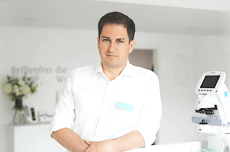
CHIEF REFRACTIVE SURGEON AT SWISSLASER KATOWICE

Dr. med. univ. (Vienna), FEBO Victor Derhartunian

A doctor by vocation, Swisslaser’s chief surgeon in Warsaw and Krakow, with more than 10 years of experience in performing refractive surgery procedures. He studied medicine at the University of Vienna, and learned the profession of ophthalmology from the pioneers of laser surgery: prof. Dr. n. med. Thomas Kohnen (University Clinic of Ophthalmology in Frankfurt) and Prof. Dr. n. med. and Dr. n. atr. Theo Seiler (IROC Institute in Zurich), who performed the world’s first laser vision correction.

Patients particularly praise Dr. Derhartunian’s professionalism and empathy, and appreciate his personalized approach and the thoroughness of the procedures performed.
Congenital cataract (cataracta congenita)

Congenital cat aracts (cataracta congenita) can take the form of:

  • stratified,perinuclear cataracts (cataracta zonularis, perinuclearis) – this is the most common case of congenital cataracts, the visual impairment is only partial, the cataract is less saturated in the center
  • nuclearcataract (cataracta nuclearis) – thecataract (cataracta nuclearis is located in the center of the lens
  • total cataracts (cataracta totalis) – causes secondary vision loss, strabismus and nystagmus
  • anterior andposterior capsular cataracts (cataracta capsularis anterior et posterior)
  • anterior orposterior polarcataract (cataracta polaris anterior velet posterior)
  • membranouscataract (cataracta membranacea)


Complicated cataract (Latin: cataracta complicata)

It appears as a complication after other chronic eye diseases (e.g. glaucoma, retinal detachment, pigmentary retinopathy, inflammation, high myopia) or systemic diseases (e.g. diabetes, hypoparathyroidism).

Complicated cataracts will also be called drug-induced cataracts, resulting from the adverse effects of certain drugs, such as steroids, which are used, among others. Among others. In asthma and Rheumatoid Arthritis or Crohn’s Disease, among others.

Acquired/degenerative cataract (Latin: cataracta acquisita)

This term is used to describe the opacification of the lens of the eye that occurs with age and is associated with aging and accompanying metabolic disorders – hence the popular term senile cataract. Based on the degree of opacity of the lens, we divide acquired cataracts into incipient cataracts (cataracta incipiens) – when there is only opacity at the periphery of the lens, and mature cat aracts (cataracta matura) – when the opacity of the lens is already complete. There are still intermediate stages between these two: advanced cataracts and immature cataracts. Transperitoneal cataracts, on the other hand, are already a stage in which the lens fibers gradually liquefy, causing the lens nucleus to move downward, which can further lead to irreversible blindness.

Based on the location of the opacity in the lens, there are several types of senile cataracts:

  • Cortical cataract – the opacity is localized in the superficial layers of the lens; visual acuity disturbances may be accompanied by double vision,
  • Subcapsular cranial cataract – the opacity localizes in the posterior lens capsule, develops slowly, but is often accompanied by light splitting and associated glare making it difficult to perform certain activities,
  • Nuclear cataract – the opacity is located in the lens nucleus and leads to myopia.


Symptoms of acquired cataracts

Senile cataracts appear around 50. year of age and may initially be unnoticeable to the patient – especially if he or she already has some visual impairment. What should be of concern?

  • deterioration of distance vision
  • problem with perception and color discrimination
  • better vision on cloudy days
  • seeing objects “as through a dirty glass”
  • Deterioration of self-care due to visual impairment

To diagnose cataracts, a simple visual acuity test is not enough. The doctor must perform an ophthalmoscopy, which is a viewing of the fundus of the eye. This is a non-invasive examination using a special ophthalmoscope speculum, which allows a thorough check of the changes seen in the fundus of the eye. Equally important is biomicroscopy, or slit-lamp examination. The test must be preceded by the administration of a pupil-dilating drug.

Treatment of acquired cataracts

In the early stages of cataracts, drugs can be administered in the form of drops, but their action does not always have the desired effect. In fact, the only effective way to eliminate senile cataracts is surgery to remove them. Until recently, only mature cataracts were operated on, but now ophthalmologists take the position that cataract surgery is advisable even in the early stages, as long as the visual impairment is severe enough to impede daily functioning.

Cataract phacoemulsification – a method of removing cataracts using an ultrasonic tip and implanting an artificial intraocular lens. The procedure is performed under surface anesthesia applied as a gel or drops into the conjunctival sac. In place of the removed lens, a new one is inserted – a flexible, retractable, acrylic lens that unfolds on its own in the natural lens bag and settles in the right place in the eyeball. The incision is minimal (1.5 to 3 mm). We have many types of lenses that can, by the way, correct vision defects such as myopia, hyperopia, presbyopia or astigmatism.

High-precision femtosecond laser surgery – a method that involves making micro-incisions (called ports) in the cornea and removing the cloudy lens through them and then inserting a new lens. The treatment takes 10-15 minutes.

NOTE: It is left up to the patient to decide whether cataract surgery is performed in both eyes during a single procedure, or whether some time should pass between surgery on one eye and the other. All pros and cons should be discussed with your doctor.

What implants are used during cataract surgery?

Cataract surgery with the Ziemer Z 8 femtosecond laser!

Recovery after cataract surgery

The cataract removal procedure is not complicated, and the most common complication – displacement of the newly implanted lens – is not the result of medical malpractice, but the patient’s return to normal daily activities too soon. Recommendations for the patient after cataract surgery are as follows:

  • It is advisable to avoid violent shocks, physical exertion and deep bending for about 2-3 months after surgery.
  • Follow-up visits are necessary, and the use of antibiotic-steroid drops (designed, among other things, to reduce pressure inside the eye) for 3-4 weeks.
  • It is important that the dropping of medication is done correctly: the lower eyelid should be pulled back and the gaze should be directed upward. The application should take place while the patient is lying down – afterwards, the eyelids should be closed for about a minute.
  • The surgical wound heals for about six weeks. During this time, you may have the sensation of a foreign body in the eye, but under no circumstances should the eye be rubbed or pressed.

It is estimated that complications after cataract surgery affect only 1-2% of cases. Among the most common are intraocular hemorrhage and bacterial intraocular inflammation.

About 3,000 cataract surgeries are performed in Poland, per million population, and according to the WHO, untreated cataracts are one of the most significant causes of reversible vision loss.

Sign up for a casual visit and consultation where you’ll learn how we can improve your eyesight.


See how to make an appointment