Age-related macular degeneration (AMD) is an age-related degenerative disease. AMD is estimated to affect as many as 30% of people over the age of 70, but the first noticeable symptoms of the disease can be observed as early as 50. The number of patients in Poland is estimated at around 2 million people, with increasing life expectancy expected to generate a steady increase in the number of patients.
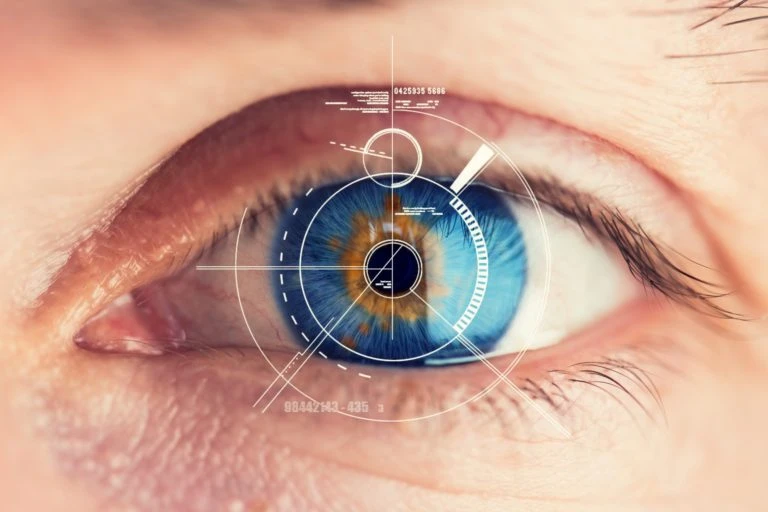
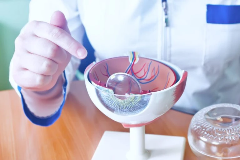
Among the first symptoms of AMD is a minimal loss of central vision, which can go unnoticed – especially if it occurs in only one eye while the other is working properly. The macula(Latin: macula lutea) is the area on the retina with the greatest visual resolution associated with the highest density of cones. The photoreceptors of the macula contain yellow pigment, hence its name.
In the dry form of AMD (non-neovascular), the disease is characterized by a slow and painless progression, so it is easy to ignore symptoms. In its course, there is a slow atrophy of photoreceptors, so-called cones, which are involved in the process of vision.
In the case of AMD in the exudative (wet, neovascular) form, significant deterioration of vision can occur rapidly, even within a few days, and in this case there is no possibility of delaying a visit to the ophthalmologist. About 10% of patients develop this form of AMD. It involves the growth of abnormal blood vessels under the central retina. From these vessels, fluid or blood can seep into the retina, causing a dark spot in the center of vision.
Classic AMD symptoms:
- reduced visual acuity (blurred central vision, or “straight ahead”),
- abnormalities in seeing straight lines,
- A bright or dark spot in the center of the visual field (known as central darkness),
- deformed shapes of objects,
- impression of less color contrast
What else should we know about both forms of macular degeneration?
The dry (non-neovascular) form is a milder form, and deterioration of vision occurs over months or even years. This form is characterized by the presence of so-called druses, or deposits of products of retinal metabolism, and so-called geographic atrophy. The aforementioned druses (deposits), deposited in the macular area, tend to merge together and cause poorer vision in low light, the impression of crooked letters or their thickening when reading. Geographic atrophy, on the other hand, is a clearly demarcated, circular or oval area of the retina in the macula that results from atrophic changes in the retinal pigment epithelial layer, photoreceptors and capillaries of the choroid of the eye. This lesion causes a slow decline in visual acuity, worse low-light vision and difficulty reading.
The exudative (wet, neovascular) form is a severe form in which vision loss occurs even within a few days. First, letters begin to ripple, and soon after, a large gray or black spot is revealed in the field of vision, making it impossible to read or see at close range. This wavy sensation is caused by the elevation of the retina, under which new pathological blood vessels with thin walls have formed, through which blood serum or blood itself oozes (hence the name: exudative). When the vessel walls rupture, blood spills under the retina and the patient sees a gray or black spot in the central visual field.
It is worth being aware that the neglected and untreated dry form of AMD can lead to the development of the disease into an exudative form, the treatment of which by intravitreal injections of anti-VEGF preparations can stop the disease process, but no longer reverse it. VEGF(Vascular Endothelial Growth Factor) is a protein considered the main factor responsible for the process of angiogenesis and the increase in blood vessel permeability; it is also called vascular permeability factor. It is the genetically increased activity of this protein that causes the aforementioned pathological blood vessels to appear under the retina.
Risk factors associated with the development of macular degeneration include:
- age
- Gender (the risk of occurrence is higher in women than in men)
- Race (people of the white race get sick more often)
- Genetic conditions (relatives of a person with AMD are 20 times more likely to have AMD compared to the general population)
- cardiovascular diseases
- diabetes
- obesity
- cigarette smoking
- drinking excessive amounts of alcohol
- Overexposure to the sun (UV radiation has a damaging effect on retinal pigment epithelial (RPE) cells, which play a key role in the proper functioning of the retinal photoreceptors)
- Poorly balanced diet (excessive consumption of foods rich in saturated fatty acids and cholesterol)
Can the onset of AMD be prevented?
If you are in the risk group, it is advisable to supplement your diet with high doses of vitamins and antioxidants. It is especially recommended to consume vitamin C (500 mg/day) and vitamin E (200-400 mg/day). It is also worth reaching for lutein, which protects against ultraviolet radiation that damages photoreceptors 6 mg of lutein per day. Natural sources of lutein are green plants: spinach, kale or broccoli.
AMD Treatment with 2RT Laser
Laser 2RT(Retinal Rejuvenation Therapy) is used to treat early stage macular degeneration. Laser 2RT is the best method to stop the development of macular degeneration before the disease takes a dangerous exudative form. The treatment aims to reduce retinal drusen and stimulate retinal structures for natural biological regeneration processes. The laser uses a light wave beam with a wavelength of 532 nm, which, combined with a short duration, leads to stimulation of the retinal pigment epithelium (RPE) and regeneration of the tissue. The procedure is painless, non-invasive, causes no thermal damage to the retina and has no side effects.
As part of the qualification for the treatment, symptoms that could make the therapy ineffective (wet form of AMD, presence of pseudodrue) should be excluded. Among the diagnostic tests needed as part of qualification are: OCT(Optical Coherent Tomography of the Eye), tonometry(intraocular pressure test), measurement of visual acuity without correction and in the best possible correction. An in-depth medical history is also necessary.
Conjunctival injections of anti-VEGF preparations
In the treatment of advanced AMD, or its exudative form, intravitreal injections (injections into the eye) of anti-VEGF preparations are used. The task of the drugs is to inhibit the growth of abnormal (pathological) blood vessels and absorb the exudate and, as a result, restore the retina to its normal position. Injections into the eye are administered under local anesthesia, so they are painless for the patient. The choice of the specific drug (VEGF inhibitor), the number of injections, the intervals between treatments and the duration of the treatment is determined by a specialist.
The following tests are performed as part of the qualification for surgery: angiography, autofluorescence, OCT. The number of AMD patients in Poland is estimated at about 1.2 million, including 10-15% of those with the exudative form.