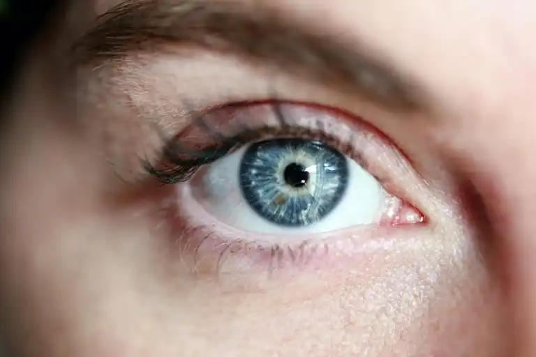
Eye Anatomy – In Brief
- The eyeball is approximately 24 mm in diameter and has three layers: outer (cornea, sclera), vascular (iris, ciliary body, choroid), and inner – the retina.
- Path of light: cornea – aqueous humor – pupil – lens – vitreous body – retina – optic nerve – visual cortex of the brain.
- The macula is a small area of the retina responsible for sharp central vision, reading, and facial recognition.
- Vision defects are an optics problem, not a retina problem: laser reshapes the cornea, and lens surgery replaces the internal lens.
The eye is one of the most fascinating and important organs of the human body, allowing us to experience the world through the sense of sight. Its complex structure resembles a precisely designed biological camera, where each element plays a crucial role in the process of vision. Understanding the architecture of the eye is essential for making informed decisions regarding vision correction and maintaining eye health throughout life.
Anatomy of the Eye – Structure of Layers and Tissues
The human eye can be compared to a perfect biological camera, approximately 24 mm in diameter and weighing about 7.5 grams. Its structure consists of three main concentric layers and numerous auxiliary structures that together form one of the most sensitive sensory organs.
Layers of the Eyeball Wall
Sclera – Outer Protective Covering
The sclera is the outer, whitish layer of the eye, composed of dense collagenous connective tissue. It covers about 85% of the eyeball’s surface and serves a protective function, maintaining the eye’s shape and providing an attachment point for the extraocular muscles. In the anterior part of the eye, the sclera transitions into the transparent cornea.
Uvea – Middle Vascular Layer
The uvea is a richly vascularized middle layer, consisting of three parts:
- Iris – the colored part of the eye with a central opening (pupil), regulating the amount of light reaching the retina
- Ciliary body – a structure containing muscles that change the curvature of the lens and produce aqueous humor
- Choroid – the layer nourishing the outer layers of the retina
Retina – Inner Neural Layer
The retina is the most important visual structure, containing photoreceptor cells (rods and cones) and neurons that process light impulses. The macula, with its fovea, is located where cones are most concentrated and is responsible for sharp central vision.
Key Refractive Structures
Cornea – The Eye’s Main Lens
The cornea is a transparent, convex structure approximately 11-12 mm in diameter, serving as the front window of the eye. It has a refractive power of about 43 diopters, making it the most important element of the eye’s optical system. Its curvature and transparency are maintained by a specific five-layered structure and the absence of blood vessels.
Lens – Variable Biological Lens
The lens is a transparent, elastic structure approximately 9-10 mm in diameter, suspended by zonular fibers. Its unique property is the ability to change its curvature through the contraction of the ciliary muscle, which enables accommodation – adapting visual acuity to different distances. The refractive power of the lens is about 15-20 diopters, depending on the state of accommodation.
Vitreous Body – Filling the Interior of the Eye
The vitreous body is a transparent, jelly-like substance filling about 80% of the eyeball’s volume. It consists of 99% water, as well as collagen and hyaluronic acid, maintaining the eye’s shape and the transparency of the optical pathway between the lens and the retina.
Accessory Structures of Vision
Anterior and Posterior Chambers
The space between the cornea and the iris (anterior chamber) and between the iris and the lens (posterior chamber) is filled with aqueous humor. This transparent fluid supplies nutrients to avascular structures and maintains proper intraocular pressure.
Optic Nerve
The optic nerve is a bundle of approximately 1.2 million nerve fibers that transmit visual information from the retina to the visual cortex of the brain. Its diameter is about 3-4 mm, and its length is about 50 mm in the orbital segment.
Physiology of Vision – How the Eye Processes Light into an Image
The process of vision is a complex sequence of events during which electromagnetic light waves are converted into nerve impulses interpreted by the brain as an image.
Path of Light Through the Eye
Stage 1: Light Refraction Light rays, passing through the cornea, undergo the greatest refraction due to the difference in refractive indices between air (1.0) and corneal tissue (1.376). The light then passes through the aqueous humor and the pupil, whose size regulates the amount of light reaching the retina.
Stage 2: Accommodation The lens automatically adjusts its curvature depending on the distance of the observed object. When looking into the distance, the ciliary muscles relax, and the lens flattens. When viewing nearby, the muscles contract, increasing the curvature of the lens.
Stage 3: Focusing on the Retina A properly functioning optical system of the eye focuses light precisely on the retina, creating an inverted and reduced image of reality.
Processing of the Visual Signal
Photoreception The retina contains two types of photoreceptor cells:
- Rods (about 120 million) – responsible for vision in low light and motion perception
- Cones (about 6 million) – enabling color vision and sharp central vision
Signal Processing Light impulses activate photopigments in photoreceptors, generating electrical signals. These signals are then processed by bipolar cells, ganglion cells, and other retinal neurons before being transmitted via the optic nerve to the brain.
Interpretation in the Brain In the visual cortex of the brain, signals are interpreted as images, taking into account color, shape, depth, motion, and other visual characteristics.
FAQ – Frequently Asked Questions
What parts make up the eye?
The eyeball has three layers: outer (cornea and sclera), middle vascular (iris, ciliary body, choroid), and inner, which is the retina. The interior is filled with aqueous humor, the lens, and the vitreous body. Additionally, there is a protective apparatus: eyelids, conjunctiva, tear film, and extraocular muscles.
What is the most important part of the eye?
It’s difficult to pinpoint one: the cornea and lens focus light, the retina converts it into nerve impulses, and the macula is responsible for sharp central vision. The optic nerve transmits the image to the brain – damage to any of these elements impairs vision.
Which part of the eye is responsible for vision defects?
Myopia, hyperopia, and astigmatism result from a mismatch between the power of the cornea and lens and the length of the eyeball. Therefore, laser vision correction reshapes the cornea, and lens surgery replaces the internal lens – in both cases, the optical system of the eye is changed, not the retina.
How many diopters does a healthy eye have?
The entire optical system of the eye has a power of about 60 diopters, of which the cornea contributes about 43 D, and the lens at rest 17–20 D. A healthy eye without a defect has a refraction of 0 D, meaning light focuses precisely on the retina.
What we do when the eye stops seeing sharply
Knowing the structure of the eye makes it easier to understand what vision correction procedures involve — laser works on the cornea, lens surgery replaces the internal lens.
- Laser Vision Correction — working on the cornea
- Lens Surgery — intraocular lens replacement
- Examinations and Diagnostics — how individual eye structures are assessed
- OCT Examination – retinal layers in cross-section
- Myopia (Nearsightedness)
- Hyperopia (Farsightedness)
- Astigmatism